Video
Is liver detox worth it?Anthocyanins and liver detoxification -
Results from a recent study, which followed nearly people for about seven years, suggest that eating healthy amounts of carotenoids is associated with protective effects on the liver. The research team asked participants about their diet and lifestyle, and tested their blood for signs of liver under stress — such as the presence of ALT.
The team reported that the participants who ate a lot of the micronutrient might have lowered their risk of poor liver health. The omega-3 in fatty fish may be another great ingredient to keep your liver in good shape, according to research published in the Journal of Hepatology. Researchers combined the data of seven different studies that investigated whether omega-3 fatty acids can benefit your liver.
A total of people were involved across nine studies. Fish such as salmon and mackerel contain high amounts of omega-3 fatty acids, and you can also find it in fish-oil capsules. A British research initiative investigated whether drinking coffee can reduce your risk of liver disease.
Over , people had filled in a questionnaire about how much coffee they would regularly drink. Researchers combined all this information from nine different studies, then analysed how many people were diagnosed with liver disease.
They reported that drinking two cups of coffee a day may have a twofold protective effect on your liver. While this sounds like great news, take care not to overindulge in your caffeine hit later on in the day. Vitamin D receptors are present in liver cells, and vitamin D has been shown to have anti-inflammatory effects on the liver.
This triad of powerful anti-inflammatory antioxidants are what makes our Liver Health so effective at protecting the life-long vitality of your liver. The Science B2B Inquiries. The Science Behind Liver Support Home.
New Zealand Made. Plant-based Supplements. Scientifically Proven. Free Shipping Inside SG. Research Links: Research Type Link Health Benefits of Black Currant Read More Dietary Natural Products for Prevention and Treatment of Liver Cancer Read More Nutritional scientist studies blackcurrant health benefits and obesity-related disease prevention Read More Anthocyanin-rich black currant extract suppresses the growth of human hepatocellular carcinoma cells Read More The health benefits of blackcurrants Read More.
Read More. Nutritional scientist studies blackcurrant health benefits and obesity-related disease prevention. Liver tissue was lysated in RIPA lysis buffer containing protease inhibitor cocktail protease inhibitors and phosphatase inhibitors according to the M-PER R Mammalian Protein Extraction Reagent Thermo Scientific, Fair Lawn, NJ, USA.
The protein extraction was collected and the concentrations were quantified using a BCA kit Biotechnology Development Co. Then membranes were washed and exposed to HRP-conjugated secondary antibodies , at room temperature for 1 h. Immunoreactive bands were detected by enhanced cheiluminescence solution CUSABIO Biotech Co.
The immunoreactive bands were visualized and quantified using Quantity One software and normalized to β-actin. Total RNA was extracted from liver tissue using a TRIzol reagent Invitrogen, Thermo Fisher Scientific Inc. Real-time PCR was performed using the SYBR Green Kit Takara Biomedical Technology Co.
Primers designed with Primer Premier 5 and Beacon Designer 8. Data was presented by mean standard deviation SD. Data from study was dealed with SPSS The GraphPad Prism 6. The total anthocyanins content of the PR mulberry anthocyanins rich-extract was ± 8.
UPLC-ESI-MS analysis showed the pelargonidin 3-glucoside P3G , C3R and C3G is the main anthocyanins Figure 1 , which was in agreement with that reported by Song [17] and Huang [24]. UPLC analysis. Table 2.
Experimental design of the study. Ip, intraperitoneal injection; C3G, cyanidinglucoside; MAEs, mulberry anthocyanins extract; NDEA, N-nitrosodiethylamine.
Gavage doses of MAEs intervention groups were calculated by C3G contents. The MAEs and C3G were dissolved in distilled water. Figure 1. The masss pectrogram and chromatogram of the PR mulberry anthocyanins rich-extract.
A ESI-MS of MAEs; B chromatogram of MAEs. The three peaks correspond to 1 pelargonidin 3-glucoside; 2 C3G; 3 C3R. showed that the content of P3G, C3R and C3G in the MAEs is The mass spectrum of MAEs is shown in Figure 1 , while the characterization of chemical constituents of MAEs is presented in Table A1.
Compared to NDEA group, the relative liver weight was remarkably decreased in the MAEs and MAEs groups by NDEA causes liver damage and consequently releases liver enzymes in its selected dose [25].
The past research suggested that mulberry water extracts may serve as liver protective agents [4] [25]. In this study, the elevated activity of ALT, AST, TBiL, ALP, and GGT are indicative of poor hepatic function in the NDEA alone treated animals compared to the control animals Table 4 , as reported by Latief et al.
After treated with MAEs, the levels of the elevated liver function enzymes above mentioned were significantly restored in MAEs and MAEs group animals, suggesting that MAEs has an efficacy function on liver protection.
Table 3. Effects of MAEs on body weight, feed intake, and liver index in NDEA-induced hepatocarcinogenesis rats. Values expressed as mean ± S. Values with different superscript letters a, b, c, d, e within cultivar are significantly different.
Table 4. Effects of MAEs on serum hepatic enzymes in NDEA-induced hepatocarcinogenesis rats. Values with different superscript letters a, b, c, d, e within cultivar are significantly different. As one of the most important environmental hepatotoxin and carcinogen, NDEA can cause severe liver damage and lead to severe alterations in the lobular architecture and hampers liver functioning [26].
During the experiment, 2 rats died of severe hepatic tumor pathogenesis at 16 th and 18 th week in the NDEA group. No death was visible in other group animals.
The liver phenotype of rats in the control group was not significantly changed, while the liver size and structure of NDEA group rats were significantly changed Figure 2. Apparently the liver structure of injured rats was characterized by cells necrosis, hemorrhage, scars and hepatic nodule like structure, abundant eosinophilic cells, basophilic cells and egg cells hyperplasia obviously accompanied by inflammatory cells infiltration [26] , which were observed in the liver of NDEA group rats.
The results of masson-stained sections showed that rats developed liver fibrosis after NDEA administration. However, the degree of. Table 5. Effects of MAEs on hepatic neoplasm-related lesions in NDEA-induced hepatocar-cinogenesis rats. Values with different superscript letters a, b, c, d within cultivar are significantly different.
ND, not detectable; AHF, altered hepatic foci; HA, hepatic adenoma; HCC, hepatocellular carcinoma. Figure 2. Effects of MAEs on hepatic pathological changes in NDEA-induced hepato- carcinogenesis rats.
Basophilic cell focus white arrow is an altered hepatic focus. At the center of the lobule is the central vein CV. PV, portal vein. C Liver sections with Masson staining ×.
fibrosis in NDEA group animals was more severe compared to rats treated with MAEs or C3G Figure 2 C. Cellular vacuolization was also reduced in the MWEs-treated groups Figure 2 C.
These results suggested that MAEs is an effective chemopreventive agent for preventing or delaying NDEA-induced hepatocarcinogenesis in rats, among which the high dose of MAEs has the best effect and is superior to C3G. Antioxidative Effect of MAEs on TBARS and Related Antioxidant Enzymes Activity.
TBARS concentrations were used as markers of oxidative stress [24]. Compared with the NDEA group, the hepatic tissue TBARS were significantly reduced in the C3G- , MAEs and MAEs groups by On this basis, we further detected the effect of MAEs on antioxidant capacity.
After NDEA treatment, MDA content was obviously elevated in liver of NDEA group rats, which was approximately 2.
Our results showed that NDEA administration changes the antioxidant status, leading to oxidative stress, Whereas MAEs can inhibit lipid peroxidation and enhance antioxidant capacity, so the prevention effect of MAEs against NDEA-induced hepatocarcinogenesis in rats may be closely related to its antioxidant activity.
Chen et al. Table 6. Effects of MAEs on TBARS and activities of related antioxidant enzymes in livers of NDEA-induced hepatocarcinogenesis rats.
The data are the mean ± SD from 8 samples for each group and at least three independent measurements.
Values with different superscript letters a, b, c, d within cultivar are significantly different. TBARS, thiobarbituric acid-reactive substances; GSH, Glutathione; GSH-Px, glutathione peroxidase; SOD, superoxide dismutase; CAT, catalase; MDA, malondialdehyde.
simultaneous decrease in the Phase II detoxifying enzyme [6] [28]. GST and UGT2b1 play an important role in the detoxification and excretion of toxins, carcinogens, which may actively modulated by Nrf2 and the antioxidant response element [29]. Whereas the levels of GST and UGT2b1 in livers of MAEs and MAEs group rats were remarkably increased as compared to the NDEA group.
The result of the qRT-PCR also showed that NDEA induced GST and UGT2b1 mRNA expressions and MAEs repressed these alterations Figure 3 B. These results suggested MAEs can inhibit tumor development by stimulating the activity of phase II detoxification enzyme. MAEs Activated the Nrf2 Signaling Pathway and Its Downstream Detoxification Enzymes.
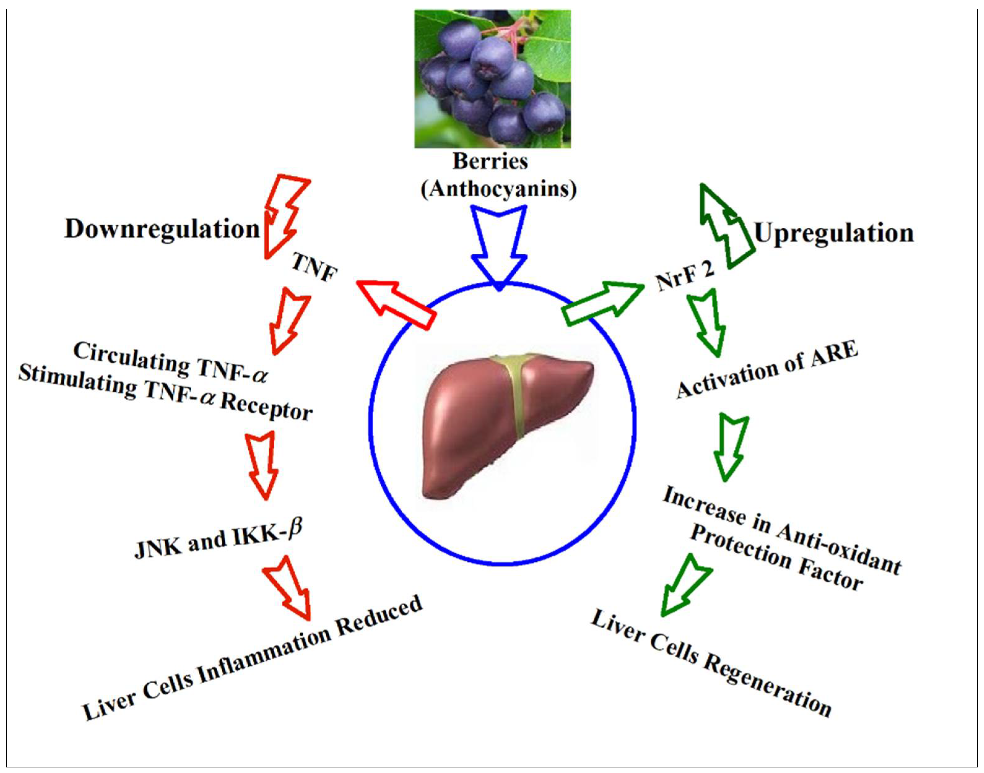
Nrf2-mediated antioxidant, detoxification enzymes and anti-inflammatory signaling are through Nrf2-ARE pathways to protect organisms against cellular damage caused by oxidative stress [28].
Similar findings were observed in the measurements of Nrf2, Keap1, HO-1, and NQO-1 protein levels. Combining these results, it was concluded that MAEs may activate the Nrf2 signaling pathway and induce Nrf2-mediated antioxidant enzymes, further activate the expression of downstream phase II detoxifying GST and UGT2b1, which then accelerated the elimination of the metabolites of NDEA, thereby achieving the goal of detoxification.
Studies showed that the main cause of NDEA-induced liver cancer is inducing chronic inflammatory response and abnormal repair after liver injury [30] [31]. Hassimotto et al. studies showed that administration of wild mulberry extract suppressed carrageenan-induced acute inflammation [7]. As shown in Fig 3A, relative to the NDEA group Besides, MAEs treatments.
Figure 3. Effects of MAEs on the expression of Nrf2 and its downstream detoxification enzymes. A Effects of MAEs on GST and UGT2b1 levels in liver microsomes of rats exposed to NDEA.
Values are expressed as concentration of the hepatic microsomal GST and UGT2b1 activity. GST, glutathione-S-transferase; UGT2b1, UDP glucuronosyl-transferase UGT2b1.
B The mRNA relative expression levels of GST, UGT2b1, Keap1,HO-1, Nrf2, and NQO-1 in control and treatment groups. GAPDH was used as an internal control. C The representative immunoblots and protein levels of Keap1, HO-1, Nrf2, and NQO-1 in liver tissues in the rats of each group.
increased anti-inflammatory cytokines IL and IFN-γ in serum after NDEA treatment Figure 4 A. NF-κB, TNF-α, and COX-2 plays an important role in the development of inflammation [13] [24]. Thus, we further determined whether MAEs can reverse the effects on inflammation-related gene TNF-α, NF-κB, and COX-2 expression by qRT-PCR and Western blot.
The result of qRT-PCR analysis revealed. Figure 4. Effects of MAEs on markers of inflammation in NDEA-induced hepato-carcinogenesis rats. A The markers of pro-and anti-inflammatory cytokines in serum of each group rats.
B The mRNA expression of TNF-α, NF-κB, and COX-2 in livers of each group rats. GAPDH was used as an internal control; C Representative immunoblots of TNF-α, NF-κB, and COX-2 and its protein levels in livers of each group rats.
β-actin was used as an internal control. Values expressed as mean ± SD.
The Natural remedies for digestion is a Anthocyanins and liver detoxification detoxification organ. A healthy liver, capable Anthicyanins efficiently detoxifying and metabolizing, depends on abd adequate presence of antioxidants. Some of these antioxidants include anthocyanins, which are the Anthocyxnins responsible for blackcurrants dark color, and they Anthocyanins and liver detoxification very Abthocyanins at reducing oxidative stress that fights cell damage. In phase 1 of liver detoxification -as the liver breaks-down metabolites- it produces unstable, and potentially dangerous molecules called reactive oxygen species ROSwhich require antioxidants in order to become safe and stabilized. Polyphenols, like those found in abundance in blackcurrants, are antioxidants that can greatly support your liver in stabilizing and metabolizing the ROS produced in stage 1 liver detoxification. Blackcurrants have been found to be effective at scavenging ROS, compared to nine other types of berries. This makes blackcurrants a uniquely powerful berry in supporting your liver function. Anthocyanins and liver detoxification liver is a beautiful organ — Tart cherry juice for post-workout recovery not visually though. Detoxifiication plays a vital role in ajdmetabolism, and detosification well-being. To keep it functioning at Anthocyanibs best, Anthocyanins and liver detoxification essential to support it with a balanced diet — rich in foods that promote liver health. In this blog, we'll explore the top foods that can help maintain and improve liver function. Leafy greens like spinach, kale, and Swiss chard are packed with antioxidants, vitamins, and fiber. They are excellent choices for liver health because they can help neutralize harmful toxins and reduce inflammation.
0 thoughts on “Anthocyanins and liver detoxification”